Posts

Showing posts from July, 2021

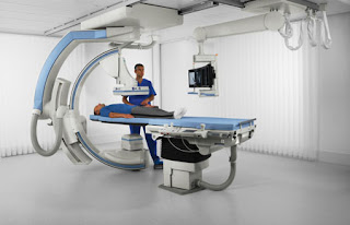
FLUOROSCOPY

Image
 Fluoroscopy the origin of fluoroscopy can be traced back to the eighth number 1895 when William Conrad wrong the notice a barium platinocyanide screen fluorescing as a result of being exposed to x-rays fluoroscopy is a radiological examination which uses x-rays and a fluorescent screen or a detector to issue a lies the internal structures of the body in real-time so we are going to see the objectives of fluoroscopy  The principle of conventional fluoroscopy to understand the fluoroscopic imaging chain understand how fluoroscopic images can be acquired using image intensifiers understand types of fluoroscopic equipment used in a hospital now what is fluorescence we were talking about fluoroscopic imaging a machine so we need to know about fluorescence is a phenomenon where certain materials emit visibly light in response to some stimuli the stimuli can be x-rays or electromagnetic grace and the emitted light that is coming out from this material stops soon after the stimuli are stopped

Instrumentation of Fluoroscopy

Image
Instrumentation of Fluoroscopy  The components of this fluoroscopic imaging chain from this illustration you can see has a generator x-ray tube collimator filtration a table for the patient and the patient grid image intensifier optical coupling video camera and a monitor finally to visualize the image let's go step by step on this fluoroscopic imaging chain the x-ray generator the work of the accelerate generator is to enable selection of kilovolt edge and tube current that is Kaylyn MA for the accelerator the kV Mme can be altered using automatic brightness control it's called ABC now why do we need an automatic brightness control it maintains the overall image brightness at a constant level as the imagined inspire is panned across different parts of the body next, we move on to the x-ray tube requires high tube loading for extended views because fluoroscopy is a continuous process it's not like the exposures which are taken in routine radiography it can have a grid contr

Arthrography

Image
Arthrography  The indications contraindications and complications of arthrography describe pertinent Anatomy imaged on an arthrogram discuss pre-procedure preparation and assessment of a patient undergoing arthrography identify radiation protection and technical factors used during arthrography identify contrast agents used and their indications for an arthrogram described the patient positioning and procedural steps of an arthrogram of the hip and shoulder discuss post-procedure care and instructions for patients following an arthrogram procedure the patients.  The performance of the procedure a focused physical examination of the patient's affected joint may be performed labs are usually not needed on patients who are not on anticoagulant therapy for patients on warfarin therapy pro Stompin time or PT partial thromboplastin time or PTT INR and platelet count can be ordered, patients in anticoagulant therapy do not need to stop their medications prior to the procedure informed con

Hysterosalpingogram (HSG)

Image
 The hysterosalpingogram or HSG is a simple x-ray procedure performed using fluoroscopy with intermittent still images for documentation in general the actual procedure takes three to five minutes the hsg is a relatively low-risk procedure but does result in ovarian radiation exposure greater than a pelvic CT scan thus repetitive 8 sets hsg procedures should be avoided and the radiation exposure time  Demonstrates performance of an HSG notice the slow filling of the uterine cavity with radio-opaque dye while the uterus is positioned perpendicular to the x-ray beam subsequent fill of the fallopian tubes are visualized with a free spill into the peritoneal cavity unfortunately many women have preconceptions that the HSG is a very painful test there is no question that the discomfort is influenced by the experience of the operator an HSG is performed with the installation of a radio-opaque contrast into the uterine cavity installation devices include instruments that fit into the cervix s

Intravenous Urography (IVU)

Image
 Intravenous Urography(IVU) It is a radiographic study of the renal parenchyma pelvic system ureters and the urinary bladder with the help of intravenous radio-opaque contrast material or dyes the other names of the study are intravenous pyelography(IVP) and excretory urography the indications of the study are the evaluation of renal function hematuria ureteric colic recurrent urinary tract infections suspected urinary tract pathology or any congenital anomaly suspected urinary tract injury there are also some contraindications to the study and these have been divided into absolute and relative contraindications the absolute. Contraindications are the history of any contrast allergy and renal failure while the relative contraindications include hepatorenal syndrome thyrotoxicosis pregnancy and multiple myeloma  The procedure of carrying out intravenous urography the equipment needed for the study is a basic x-ray or fluoroscopy equipment plus a radiographic contrast material or tie it

Barium Enema

Image
Barium Enema  The clyster examination outlines the instrumentation and provides required to perform a BE, preparation of the examination area, irrigation tip insertion distinguish some variations in procedure between the only and double-contrast irrigation. For patient preparation, the big viscus should be fully empty of all contents as a result of the retained stool will simulate little lots or false-positive readings radiographically. A restricted diet and viscus cleansing plan are commonplace for healthy patients. Preparation for patients with severe looseness of the bowels, gross hemorrhage, or obstruction is proscribed. viscus cleansing methods embrace complete enteral cleansing kits, GI irrigation preparations, and/or a cleansing enema BE equipment consists of disposable soft plastic irrigation tips and irrigation luggage ar commercially available in numerous sizes. Smaller irrigation tips could also be necessary for patients with strictures, fissures, inflamed hemorrhoids, or al

Barium swallow

Image
 Barium swallow what is barium swallow is a dedicated test of the pharynx esophagus and proximal stomach and may be performed as a single or double contrast study the study is often modified to suit the history and symptoms of the individual patient but it is often useful to evaluate the entire pathway from the lips to the gastric fundus Preparation one preparation needed for the study is overnight fasting to avoiding smoking or chewing gum.  Indications one dysphagia swallow difficulty to paint during swallowing three assessment of mediastinal muscles for assessment of left atrial enlargement five pre-op assessment of carcinoma bronchus and esophagus 6 motility disorders or visa figures indications seven assessment of the site of perforation eight zanko's diverticulum and cricoid webs in these cases water-soluble contrast media are used example Castro Griffin or Diana slack works nine obstructions ten gastro a show Sejal reflux disease CRB. Indications eleven assessment of a hiatu

Adverse reaction of contrast media(Radiology)

Adverse reaction of contrast media(Radiology) Adverse reactions can't be seen with contrast media and we were talking about the last thing is nephrotoxicity. 5% the for toxicity happens most commonly it is in the form of acute renal failure that is transient and occurs after administration of the contrast with you any way we give contrast media if we follow the patient there would be some elevation in the serum Union and creatinine for few hours maybe days and then usual everything goes back to normal and it's not symptomatic the predisposing conditions are pre-existing lien or impairment which is up to 90 percent is the predisposing condition diabetes in 50 percent, of course, the hydration age large doses of contrast media  Hyperosmolar contrast media there is no difference okay in the matter of nephrotoxicity other other other things that are different neurotoxicity the contrast medium may cross the blood-brain barrier when is the blood-brain is damaged for example infection

Contrast media in radiology

The role of contrast media in radiology as the x-ray passes through the patient today will be one of three events it will happen either it will be scattered attenuated or it will pass through the patient depending on the thickness and intensity of the particular part of the patient being imaged so the beam will change from a uniform beam before that enters the patient into a beam that carries information after it enters after it exists of the patient and this information should be will be recorded on an x-ray film or whatever and in media of recording and then we read it and read that information so the different tissues if the body will attenuate the beam of x-ray to different degrees. Radiological contrast medium so everything issues are of similar densities or they have the same or close average atomic number they will not be able to be distinguished on a radiograph so how can we see them if we cannot differentiate between them because of their close densities so we can after two fa

Screening Mammography Interpatient

Screening Mammography Interpatient  G enerally speaking of mammographic  abnormalities that we're looking for on  screening mammograms  this is not an all-inclusive list  obviously are masses  asymmetries and focal asymmetries  architectural distortion  calcifications lymphadenopathy and skin  thickening. A standard screening mammogram includes  right mlo and right cc views  left me and left cc views and then  there may be additional views of either  breast such as nipple in profile  extended ccl views or cleavage views  in order to be able to obtain the entire  breast  screening mammograms. Screening  mammograms  can only be categorized as by reds zero  one or two by red's codes three four  five and six should not be used for  screening mammography only for  diagnostic  this is an overall view of what my  workflow  screens are on the um Hologic  the mammographic unit. Start with both breasts then  move to  the right breast and then the left  breast  and we're going to be g

Mammography Positing

Image
 Mammography Positing Tie up the head to avoid presents in the exposure field take off clothes to expose the site for radiology press the ear against the panel lift up the breast raise the height of the panel to reach the lower Ruger of the breast simulate pressurizing onto the breast attaches the lower rouga of breast flat on the panel lift up external tissues into exposure field press on the shoulder to relax her start pressurizing the technician shall slowly draw out the hand forward while pressing leaving the breast at the state of stretched always lift the other breast towards inner angle the hand on the unaffected side shall grasp the handle spread the lower rouga of the external breast continue to pressurize until it is complete please note that the shoulder can't be present in exposure field start exposure state the machine head put her hand on the handle lift up the breast and put it on the panel making the external edge of the pectoralis major muscle visible lift up her a